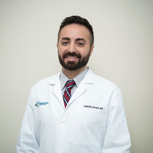
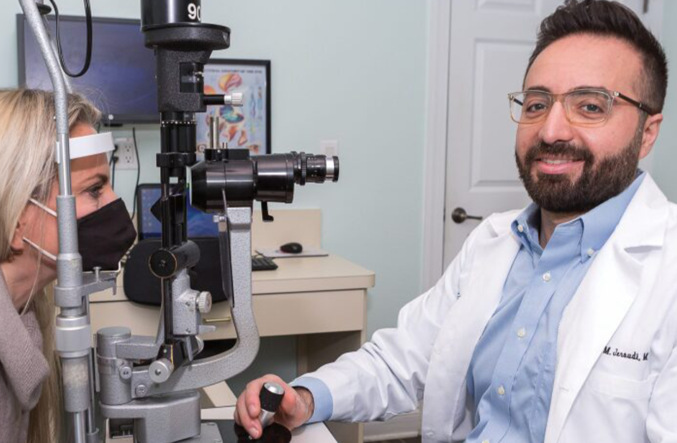
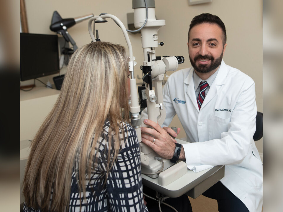
Dr. Jeroudi can typically diagnose CRVO during a physical eye exam.
The eyes are considered the “windows to the soul.” They are also the windows to the world. Individuals receive about 80 percent of the information about their surroundings through their vision.
Unfortunately, there are many eye disorders that can decrease vision. One of them is a type of stroke in the eye called a central retinal vein occlusion, or CRVO.
“The retina is fed by blood vessels that emanate from the optic nerve,” explains Abdallah M. Jeroudi, MD, a board-certified, fellowship-trained retina specialist at Florida Retina Institute. “There are arteries that carry blood to the retina and veins that collect blood from the far reaches of the retina and drain in the back of the optic nerve. CRVO is an obstruction at the main trunk of the major retinal vein, the central vein, right at the nerve.
“To understand CRVO, think of those characters in old cartoons who step really hard on a firehose and the hose starts to bubble up behind their feet and get bigger. Imagine the excess pressure in the vein because it is unable to drain. That makes the blood vessels appear wiggly and causes three things to occur that can harm vision.”
For one, the pressure within the vessels causes blood to begin leaking out, resulting in flame-shaped hemorrhages in the retina. Second, fluid leaks out of the vessels, which leads to swelling in the retina that can reduce vision. Third, when blood isn’t draining, fresh blood can’t get into the retina efficiently. The lack of fresh blood to nourish the retina causes a low-oxygen injury, which can result in a loss of vision.
“CRVO usually follows a course of blood vessel damage called Virchow’s triad,” Dr. Jeroudi discloses. “The first component of the triad is the presence of certain risk factors. The primary risk factor is age. Ninety percent of CRVO cases occur in patients older than 55. High blood pressure, high cholesterol and diabetes are also risk factors.”
High blood pressure and high cholesterol can increase pressure within the eyes. Consistently high blood-sugar levels associated with diabetes damage blood vessels throughout the body, especially the tiny vessels in the eyes.
“The second aspect of Virchow’s triad is stasis, which means blood flow in the eye is not ideal because of blood vessel damage,” Dr. Jeroudi continues. “The third component is hypercoagulability. This is when the blood is a bit too thick because there’s extra stuff in it, which increases the risk for clots to form.”
Hypercoagulability can occur if there are too many cells in the blood, a condition called polycythemia vera. There may also be an excess of antibody proteins in the blood. That can occur with conditions such as multiple myeloma, a cancer of the blood’s plasma cells, and a type of non-Hodgkin’s lymphoma called Waldenström’s macroglobulinemia.
Making the Diagnosis
The primary symptom of CRVO is a sudden, painless loss of vision, Dr. Jeroudi reports. The vision loss may be mild or severe. In addition, the eye may be red and, in advanced, end-stage cases, the individual may be sensitive to light.
“Typically, a physical exam is all that is needed to make the diagnosis of CRVO,” the retina specialist says. “We look inside the eye for typical findings, such as very dilated and tortuous blood vessels. We can also see the flame-shaped hemorrhages throughout the retina.”
During an evaluation, Dr. Jeroudi also looks for something called cotton wool spots. These are retinal manifestations of other disorders, such as diabetes, systemic high blood pressure or even HIV.
“We may use an imaging tool called optical coherence tomography,” Dr. Jeroudi reveals. “OCT uses a camera and a special light to create cross-sectional images of the retina. It shows us many of those telltale signs of retinal vein occlusion.
“If there’s a concern that the patient’s problem may be something other than CRVO, we may use fluorescein angiography. During this test, we inject a plant-based dye called fluorescein into the bloodstream. It travels to the heart, which pumps it throughout the body and into the eyes. We use a special camera to view how the blood is flowing through the eye and can look for an occlusion.”
Treatment Goals
Treatment for CRVO focuses on relieving the swelling in the retina, which leads to blurry vision. The most effective way to stop the blood vessels from leaking fluid is by injecting an anti-vascular endothelial growth factor (anti-VEGF) into the eye. Vascular endothelial growth factor (VEGF) stimulates the formation of blood vessels, which with CRVO can be unstable and leak.
“Anti-VEGF medications help to stop the blood vessels from leaking, shrink the swelling in the retina and improve the patient’s vision,” Dr. Jeroudi notes. “Initially, patients receive injections about every four weeks. As we gauge their response to the treatment, we try to extend the interval between injections.
“There are other eye injections that may be used for this condition. These include steroid injections, although those tend to be a second-line treatment. Steroids help to tighten the blood vessels, which reduces leaking and swelling. We may inject a steroid suspension. There is also a steroid implant that can be placed in the eye.”
Treatment for CRVO is generally effective at improving vision, although success depends on the patients’ vision.
“If their vision is 20/60 or better, the prognosis for recovery is quite good,” Dr. Jeroudi states. “We cannot get their vision perfect, but at least the patients will have decent vision. If patients present with vision that is worse than 20/60 but better than 20/200, the results are variable. It could go either way. If their vision is worse than 20/200, the patient will probably have to live with the vision they have.”
Prompt treatment of CRVO is necessary to avoid serious complications, such as the growth of blood vessels on the iris, the colored part of the eye.
“This condition can be rapidly blinding,” Dr. Jeroudi warns. “Blood vessels on the iris contribute to high pressure inside the eye. Sometimes, the pressure shoots up very high, causing one of the worst pains someone can experience.
“To help prevent CRVO, we ask patients to control their risk factors. This includes maintaining a healthy blood pressure and cholesterol level, as well as managing their diabetes and any coexisting blood clotting disorders.”


Leave a Reply