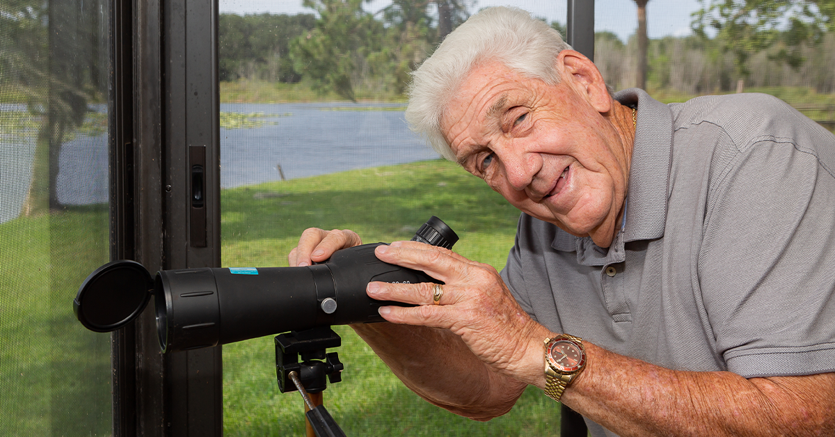

Jordan Pysz / ifoundmydoctor.com
Leon was diagnosed with aggressive wet AMD in his right eye, but after undergoing medication injections in the eye the swelling in the retina has gone down and his vision has greatly improved.
In October 1962, a US spy plane secretly took photos of nuclear missile sites being constructed by the Soviet Union on Cuba, 90 miles from Florida. The result was a tense, 13-day standoff between the world superpowers that nearly triggered nuclear war.
In response to a stout blockade of US ships that surrounded and quarantined the Caribbean island, the Soviets ultimately agreed to remove its missiles already in Cuba. The incident became known as the Cuban Missile Crisis.
Leon Norregaard had a front row seat for the drama as a member of the US Coast Guard patrolling the waters around Cuba. He helped secure waters closer to home as well.
“I went with the Coast Guard to Cuba during the crisis, but I primarily worked on the Mississippi River, the Lower Mississippi River and the Ohio River,” Leon recounts. “I also spent a couple of years in the Federal Building in St. Louis doing office work.
“I chose the Coast Guard because you have to sleep on the ground in the Army, and I don’t sleep very well on the ground. I was an Eagle Scout, so I had done enough of that while I was in the Scouts. Also, the lady at the draft board was a friend of the family. She called me and said, If you don’t enlist soon, we’re going to draft you.”
Leon served four years in the Coast Guard and another two years with the Coast Guard Reserve. After he was discharged, he began what became a lengthy career in the corrugated box industry.
“I worked 20 years in manufacturing doing various jobs, from being a baler compressing leftover box material into small bundles to running a plant,” Leon relates. “After that, I went into sales in Florida for about 15 years. Then I started my own box plant in Jacksonville with a couple other fellows. I retired in 2006.”
Even in retirement, the 82-year-old Illinois native doesn’t let grass grow under his feet. He stays active with his favorite seasonal pastimes. He also serves as landlord and caretaker for a group of rental properties owned by him and his wife. Maintaining the properties takes a great deal of time, which he gives up willingly despite the challenges.
“We live on Lake Weir on Bird Island, and we go out on the boat and play on the lake during the summertime,” Leon details. “In the wintertime, we go on cruises and go to shows at the (Ocala) Civic Theatre. Just the normal stuff. I also work on the rental properties; we’ve got eight or nine of them right now. Working on them isn’t much fun.”
It became even less enjoyable when a troubling eye issue emerged.
“I woke up one morning a couple of years ago with a (blind spot) in the center of my right eye,” Leon describes. “As the day went on, that blind spot started to fade and I would see normally again, but it continued to appear in the mornings.
Leon says he “eventually adjusted” to the problem, but during a regular appointment he mentioned it to his eye doctor with the Department of Veterans Affairs.
“She said, I’m going to send you to Florida Retina Institute to have it checked.”
About AMD
At Florida Retina Institute, which has offices in Lady Lake, Mount Dora, Clermont and other locations across Florida, Leon met with Alexander C. Barnes, MD, a board-certified, fellowship-trained retina specialist.
In examining Leon, Dr. Barnes diagnosed the blind spot as a symptom of wet macular degeneration, a condition primarily associated with age that affects millions of people. It is also referred to as age-related macular degeneration, or AMD.
“Besides age, another major risk factor is race,” Dr. Barnes informs. “Folks who are Caucasian have a higher risk for AMD, as do those who have a strong family history of the disorder. Smoking can increase the risk as well.”
There are two types of AMD: dry and wet. About 85 percent of patients diagnosed with the disorder have the dry form, which occurs due to natural degenerative changes to the retina.
The retina is the layer of nerve tissue that lines the back of the eye. It converts light rays into electrical signals and sends those signals to the brain, where they are interpreted as visual images.
Dry AMD, which typically causes blurry or distorted vision, begins with the formation of small protein deposits called drusen under the retina.
There is no known cure for dry AMD, but its progression may be slowed with a vitamin supplement called AREDS2, which was developed by National Eye Institute researchers in a six-year clinical trial called Age-Related Eye Disease Study 2.
In rare cases, dry AMD can progress to the more serious wet form.
“With wet AMD, a network of abnormal blood vessels grows under the retina,” Dr. Barnes educates. “These vessels can leak fluid and sometimes bleed, and this can destroy much of a patient’s central vision.
“Wet AMD can drastically affect a person’s quality of life if it is allowed to progress to more advanced stages, so part of our job as retina specialists is to catch changes to the retina at an early stage to try to preserve central vision.
“In addition to blurry or distorted vision, wet AMD can also cause dark spots to form in a person’s field of vision, which is what happened with Mr. Norregaard. The good news is that the wet form can be treated, and further vision loss can be prevented if we catch it early enough.”
Dreading the Needle?
The mainstay of treatment for wet AMD are injections of medication into the eye, Dr. Barnes explains. These medications act against vascular endothelial growth factor (VEGF), a protein that promotes the growth of the abnormal blood vessels and causes them to be more destructive. Anti-VEGF medications stabilize the development of the harmful vessels.
“The good news about wet AMD today that was not true a generation or two ago is that there are now several agents available to treat the disease,” Dr. Barnes contends. “If a patient is not responding to one medication, the retina specialist can petition the insurance company to switch to another medication to see if it has a better effect.
“One of the anti-VEGF medications we use is called bevacizumab, or AVASTIN®, which many insurance companies want us to start with. There is also ranibizumab, or LUCENTIS®, and aflibercept, or EYLEA®. These medications act on a similar pathway. There is also a newer medication that acts on a slightly different pathway called faricimab, or VABYSMO™.
“There is a whole other class of medications being developed called biosimilars, which are like generics. Essentially, there are differences in the manufacturing process with these medications compared to the established anti-VEGF medications, and they tend to be less expensive. A biosimilar for LUCENTIS called CIMERLI™ is currently on the market.
“We are going to be seeing more and more of these biosimilar medications available, and we will incorporate them into our practice at Florida Retina Institute as they become available.”
Anti-VEGF medications only stay inside the eye for a certain amount of time before the body flushes them out. As a result, the injections must be given regularly. Each person with wet AMD is different, so the interval between injections is specific to each patient, says Dr Barnes.
“Typically, we begin with monthly injections,” he says. “As the disease stabilizes, we try to extend the interval between injections. When Mr. Norregaard came to us, he had pretty extensive disease, which we treated quite aggressively with injections twice monthly into his right eye.
“It took us a little while to get Mr. Norregaard’s condition under control, but it seems he has reached a steadier state. He is now receiving an injection every two months. We will try to extend the interval further as his eye allows.”
A Good Trade-Off
The mere idea of an injection into the eye can be very unsettling, even frightening. But Dr. Barnes says they are typically well-tolerated by the millions of AMD patients receiving them.
“Patients come into our office with changes to their vision, distortion or marked loss of vision, and when we tell them they need a shot in the eye it can be anxiety-provoking,” Dr. Barnes relates. “I think it helps for patients to know that many of their peers are getting these injections and tolerating them.
“Having these medications is a huge benefit to patients with wet AMD because it was not that long ago when this was truly a blinding condition. Fortunately, because of the medications, patients can lead a high quality of life with the caveat that they must come in every so often for their injections.”
The retina specialists at Florida Retina Institute make great efforts to ensure the injections are as pain-free as possible by using a combination of numbing drops and a numbing injection.
“When patients first hear the news that they need an injection into the eye, their knee-jerk reaction is to think it is going to be painful,” Dr. Barnes states. “But after the first injection, most patients are surprised pain is really not a part of the process. Most do not report pain with the injections.
“The first injection is really an emotional and psychological hurdle to get through, and every patient is different. Some people are affected more by the process than others. But most people understand the goals for treatment.
“They understand that an injection may not be the thing they look forward to most in life, but it happens one day out of the month, and it allows them to enjoy the things they want to do the other 29 days. It really is a pretty good trade-off.”
Leon is among the many who came to realize that the idea of an eye injection is a lot worse than the actual injection.
“The injections weren’t painful at all,” Leon asserts. “Dr. Barnes is a great doctor. He did a wonderful job so the shots didn’t hurt. I get a few floaters occasionally when he first injects the medication, but Dr. Barnes says those are air bubbles, and they usually go away that day or the next.”
Change of Schedule
When Leon initially visited Florida Retina Institute, Dr. Barnes examined his right eye and diagnosed wet AMD. He gave Leon an injection into his eye and told him to come back to the institute in a month. Leon complied, but his eye wasn’t much better.
“During that second visit, Dr. Barnes gave me another shot and said, Come back in two weeks. We’re going to put you on a two-week schedule for a while and see what happens,” Leon remembers. “After a few weeks of being on that schedule, we could see that the swelling in my retina was way down, so the treatment was working.
“After that, Dr. Barnes started giving me an injection every three weeks. Then we went to four weeks and eventually five. The swelling kept going down and my retina was almost back to normal, so we kept putting more time between the injections and now I’m up to once every eight weeks.
“The good news is that my retina has been staying where it’s supposed to be. The black spot that appeared in my right eye in the mornings went away after I started getting the shots, and the vision issues I was experiencing got better.”
Dr. Barnes confirms that Leon is doing well with his treatment plan.
“When we started, Mr. Norregaard had a pretty aggressive disease that was concerning for me and him as to whether we would be able to control it,” Dr. Barnes recalls. “But thanks to the new medications, when we look at the scans of his retina today, it’s almost like a totally different eye. He has responded nicely to the treatment and, hopefully, he will continue to do so.”
Leon has high regard for the retina specialist who has worked so diligently to restore his eyesight. He also thinks highly of the team at Florida Retina Institute.
“Dr. Barnes is very knowledgeable. He’s in control and knows what’s going on,” Leon raves. “He has a good bedside manner, and he always keeps me informed. And the staff is very friendly. They do a fine job as well. I would recommend Dr. Barnes and Florida Retina Institute in a heartbeat.”
Calm, Cool, Reconnected
Johanna Trowell has spent the past 18 years assisting two OB-GYNs in a private practice, where she now serves as clinical manager.
“Originally, I wanted to be a pediatrician, but life goes on and things get in the way, and I decided to become a nurse instead,” recounts Johanna, 48, a Puerto Rico native. “It’s actually been very gratifying. It required less schooling, too.
“Since the beginning of my career, I wanted to work in private practice. All nurses have their niche, and the hospital wasn’t mine. I started out working in a pediatrician’s office and stayed there for a couple of years. Then I had the opportunity to join the OB-GYN practice. I stayed all these years because I really enjoy women’s health and pregnancy.”
Recently, Johanna received a clean bill of health from her eye doctor following a routine checkup and contact lens update. Unfortunately, that prognosis changed rapidly just a few days later.
“My appointment was on Friday,” Johanna recalls. “Everything turned out great. I got a new prescription and new contacts to try. Then, two days later — that Sunday morning — I noticed that the corner of my peripheral vision in my right eye was pretty blurry, like a drop of water was in my eye.
“That was in the morning; toward afternoon, that area became quite dark. On Monday, it seemed to me as if the darkness had spread. It covered a little more of my field of vision than the day before, so I called my eye doctor and got another appointment for the next day.”
During that appointment, the optometrist examined Johanna’s right eye and performed several imaging tests. The images revealed that Johanna’s retina was detaching from the back of her eye from the top to the bottom, like peeling wallpaper.
“My doctor said, You have a detached retina. I hope you don’t have any plans to travel anytime soon,” Johanna remembers. “I said, Yes, I have a trip to Europe in three weeks. It’s a two-week vacation I’ve had it planned for many months. She looked at me and said, I’m not sure about that. Let me talk to my colleagues and get back to you.
“My doctor immediately got on the phone with Florida Retina Institute and told them about my situation. They set up an appointment for me to meet with a retina specialist at the institute the next day.
“When I got to Florida Retina Institute, we scheduled surgery to reattach my retina for the upcoming Friday. But on my way home, that Wednesday, they called me and said, Ms. Trowell, the doctor thinks you should actually have surgery sooner. He’s going to have one of his colleagues do it tomorrow. That’s when I met Dr. Cunningham.”
Scleral Buckle
Matthew A. Cunningham, MD, is a board-certified, fellowship-trained retina specialist at Florida Retina Institute. He explains that there are three types of retinal detachments: exudative, tractional, and rhegmatogenous.
An exudative detachment happens when fluid builds behind the retina and pushes it away from the back of the eye. Exudative detachments are usually secondary to inflammation or a vascular abnormality. These don’t involve a retinal tear and generally resolve once the underlying cause has been addressed.
Tractional detachments occur when scar tissue pulls or causes traction on the underlying retina. Patients may not have an associated retinal tear.
A rhegmatogenous (pronounced reg-ma-TAH-juh-nus) detachment is the most common. These typically occur as the result of a retinal tear or break and can follow a posterior vitreous detachment (PVD).
A PVD arises when the vitreous gel inside the eye begins to liquefy and pulls away from the retina. PVDs occur in about 60 percent of patients and are part of the normal aging process.
“Posterior vitreous detachments typically happen in patients in their 60s and 70s,” Dr. Cunningham observes. “However, they can also occur in younger people who are extremely nearsighted. They often lead to a retinal tear and detachment.”
Symptoms of this condition include a sudden onset of flashes of light, floaters, and sudden changes in vision. People with these symptoms are urged to immediately visit an eye care specialist.
Also, people who are extremely nearsighted should pay special attention to these types of changes in their vision because they are at a higher risk for developing a retinal tear.
“Ms. Trowell had a rhegmatogenous detachment, which required surgery to repair,” Dr. Cunningham explains.
Johanna’s surgery was moved up because the detachment had not yet reached the macula, which is the center of the retina responsible for central and color vision.
“In situations like that, where the detachment does not involve the macula, we like to repair the detachment pretty quickly, usually within a day or so,” Dr. Cunningham notes. “In Ms. Trowell’s case, we performed a scleral buckle procedure, during which we place a silicone band around the sclera, the white part of the eye.
“Once it’s in the perfect position to support the retinal tear, we suture the band onto the sclera, and it acts like a suspender around the eye globe where the retinal tear is located.”
“Because we are causing an indentation in the eye with the scleral buckle, the eyeball becomes a bit elongated,” Dr. Cunningham describes. “As a result, patients typically become more nearsighted, or myopic, which can be corrected with new glasses or a new contact lens prescription. This happened with Ms. Trowell. She needed a new prescription for her contact lenses to correct the issue.
“Other than the change in her nearsightedness, Ms. Trowell has made an excellent recovery. Her retina is completely reattached, and she’s not having any other problems with her eye at this time.”
Johanna concurs.
“I don’t have any dark patches, so I’m able to see my entire field of vision now,” she confirms. “Before surgery, half of my vision was very dark.”
Johanna says this process taught her to not take early symptoms casually.
“Exactly a week before this whole thing happened, I noticed two floaters in my right eye,” Johanna remembers. “I had floaters in the past, so I never thought anything about them being a prequel to a retinal detachment. Now I know better.”


