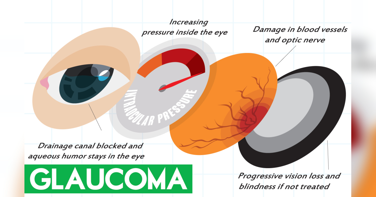
Glaucoma refers to a group of progressive eye diseases that cause vision loss by damaging the optic nerve in the back of the eye. The optic nerve is a bundle of more than 1 million nerve fibers that carry messages from the retina, where light is converted into nerve impulses to the brain. The brain interprets the impulses as visual images.

January is National Glaucoma Awareness Month. Let’s learn more about glaucoma, which affects about 3 million Americans and 80 million people worldwide. It is the second leading cause of blindness in the world, behind cataracts.
One of the main risk factors for glaucoma is increased pressure in the eyes, or intraocular pressure (IOP). High IOP damages optic nerve fibers, leading to vision loss. IOP increases when the fluid that bathes and nourishes the front of the eye, called aqueous humor, cannot drain as fast as it is created and builds up.
Fluid drains through an area called the drainage angle, located where the colored part of the eye (iris) meets the white covering over the eye (sclera). One of the main structures of the drainage angle is the trabecular meshwork, which plays an important role in draining aqueous humor from the eye and maintaining a consistent IOP.
In addition to high IOP, other risk factors for glaucoma include being older (it is more common in people ages 60 and older), being African American or Latino, having a family history of glaucoma, having diabetes (people with diabetes are twice as likely to develop glaucoma), being very nearsighted or farsighted, using corticosteroids long-term and having a thin cornea.
Anyone with a risk factor should see an eye doctor for an annual exam.
There are two main types of glaucoma:
• Open-angle glaucoma is the most common, affecting up to 90 percent of Americans with glaucoma. It develops when tiny deposits build up in the eye’s drainage canals and slowly clogs them so fluid cannot drain efficiently. It typically has no symptoms early on. As the disease progresses, peripheral (side) vision may diminish. Because the disease can get to that point without symptoms, it is often called the “silent thief of sight.”
• Angle-closure glaucoma occurs when the iris is too close to the eye’s drainage angle. It can block the drainage angle so fluid cannot drain and IOP increases. If the angle gets completely blocked, IOP increases rapidly and causes symptoms. This is called acute angle-closure glaucoma. Signs of an attack include sudden blurry vision, severe eye pain, eye redness, headache, nausea, vomiting and rainbow-colored halos around lights.
Acute angle-closure glaucoma is a medical emergency. Anyone experiencing one of these symptoms should seek help immediately.
Because it causes no symptoms early on, it is possible to have glaucoma and not know it. So, routine eye exams are important to catch glaucoma before it can severely damage the optic nerve.
A doctor may perform one or more tests to check for glaucoma. These include a dilated eye exam to view the optic nerve, a gonioscopy to examine the drainage angle, tonometry to measure IOP, optical coherence tomography (OTC) to look for changes in the optic nerve that may indicate glaucoma, a visual acuity test (eye chart test) to check for vision loss and a visual field test to check peripheral vision.
If you have glaucoma, it is important to start treatment right away. A person cannot regain any vision already lost, but treatment can help slow the progression of the disease and reduce the risk of additional vision loss. To treat glaucoma, doctors typically use medications, laser procedures or surgery.
The most common medications for glaucoma are prescription eyedrops. These work by decreasing the production of aqueous humor or increasing its flow out of the eye. More than one eyedrop may be prescribed to control IOP.
The doctor may use a laser to help fluid drain from the eye. Lasers can be used to treat open-angle and angle-closure glaucoma. A laser can open the drainage angle, make a tiny hole in the iris to let the fluid flow more freely and treat areas in the eye’s middle layer to lower fluid production.
If eyedrops and laser treatments don’t lower IOP, the doctor may suggest surgery. There are several procedures that can help drain the fluid, including trabeculectomy microsurgery, in which the doctor creates a new drainage channel.
Minimally invasive glaucoma surgery (MIGS) makes tiny openings in the trabecular meshwork and places tiny devices, such as stents and microshunts, to help drain fluid. MIGS is most often performed as an add-on procedure after cataract surgery.
Now that you’ve had a glimpse at glaucoma, share what you’ve learned this National Glaucoma Awareness Month.


Leave a Reply