
July is National Cleft and Craniofacial Disorders Awareness and Prevention Month. This observation was established to educate Americans about the cleft and craniofacial conditions that affect more than 600,000 people in the US.
Cleft and craniofacial disorders are a diverse group of abnormalities in the growth and development of the head and face. Most of these disorders develop during pregnancy and are present at birth.
There are multiple variations of cleft and craniofacial disorders, and they can be mild, moderate or severe. They often affect the physical appearance of the child, but can also impact important functions, including eating, hearing and seeing.
The exact cause of these disorders is unknown. Most physicians believe there is no single cause but multiple factors contribute to their development. These factors include a combination of genes; environmental factors, such as viruses and exposure to dangerous chemicals in the workplace; and a deficiency of the B vitamin folic acid during pregnancy.
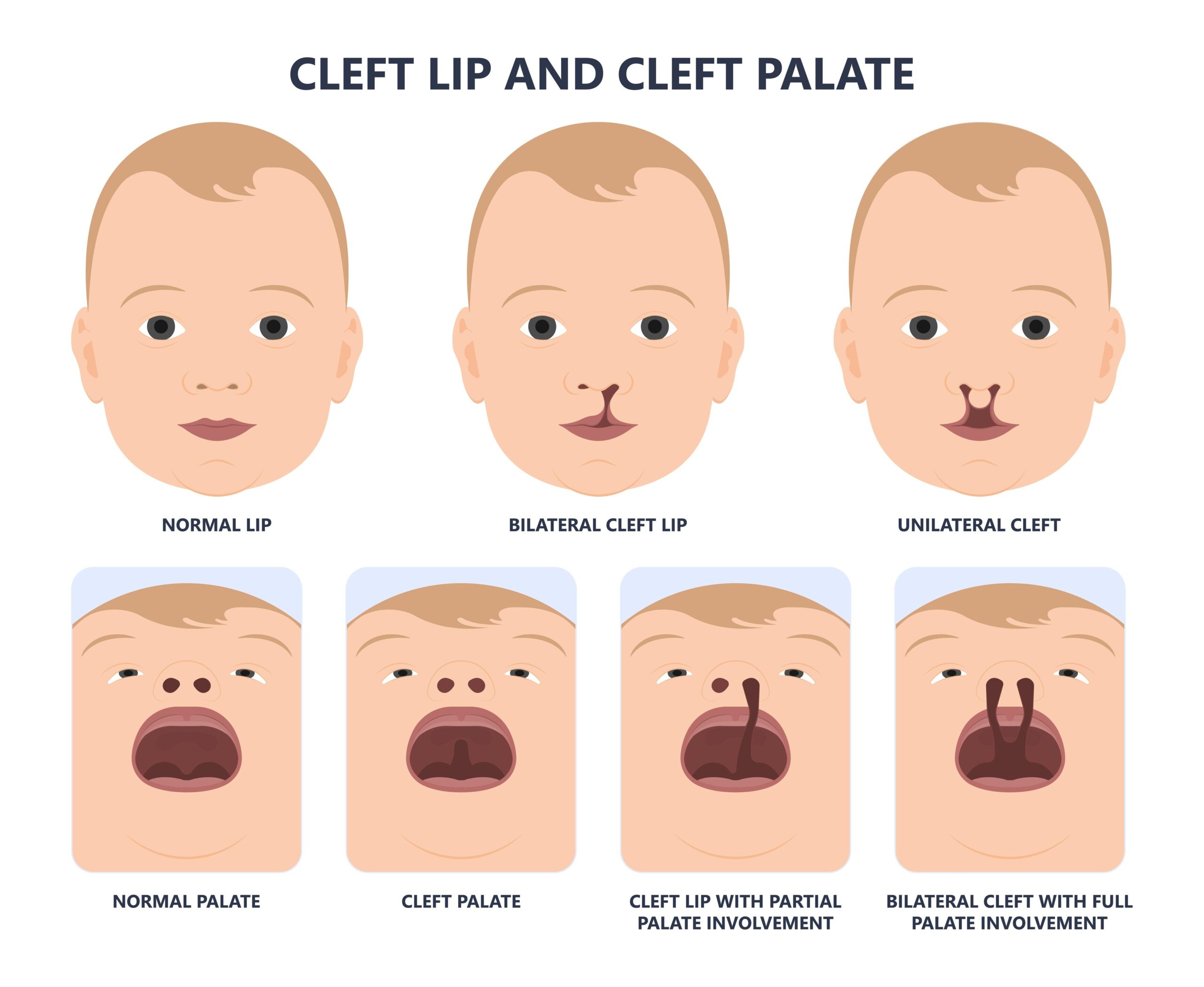
The most common of the cleft and craniofacial disorders are cleft lip and cleft palate. These conditions are the most common birth defects in the US. It’s estimated that 2,650 babies are born with a cleft palate each year in the US, and 4,440 are born with a cleft lip with or without a cleft palate.
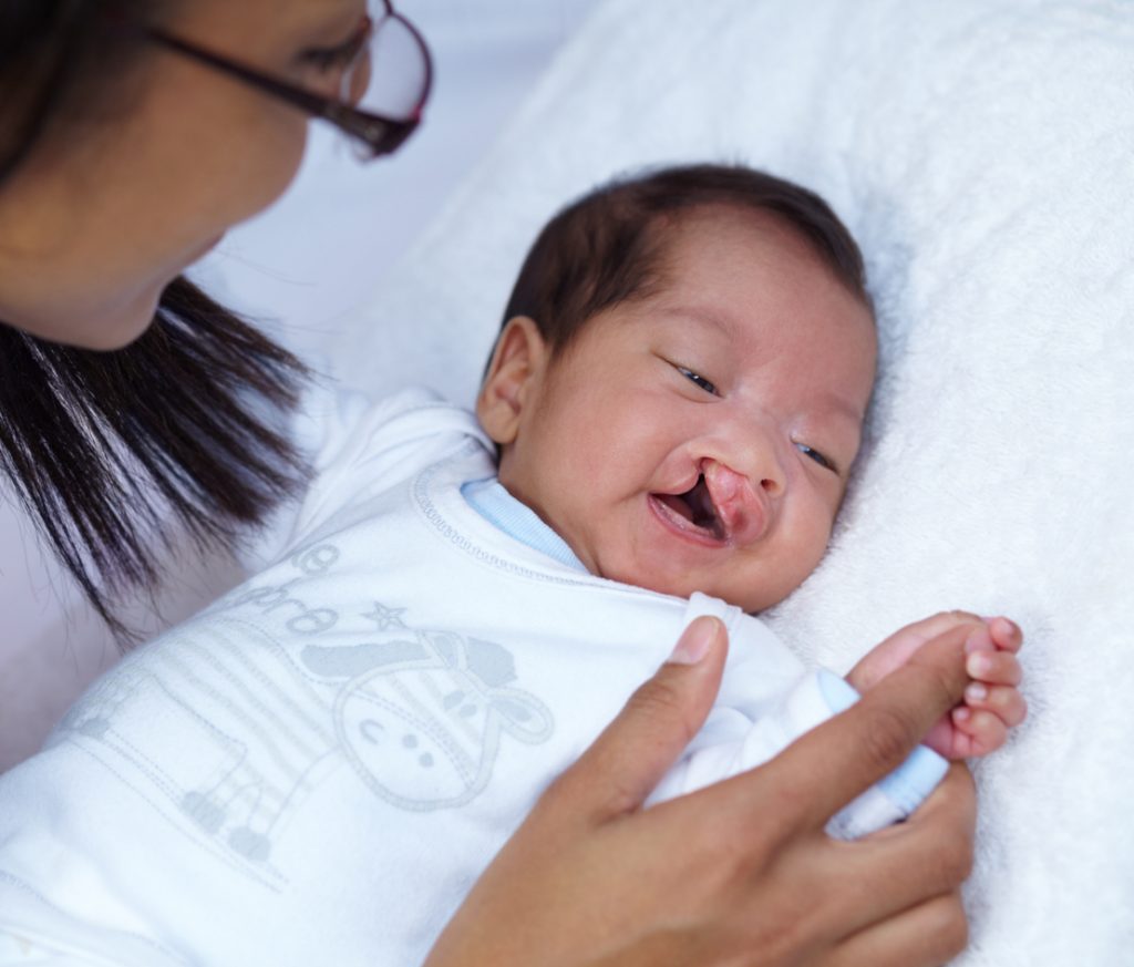
A cleft lip is a separation of the two sides of the lip. It can range in severity from a small split in the lip to a large opening that goes from the lip up through the nose. A cleft palate is an opening in the roof of the mouth. These disorders occur because the two sides did not fuse during development. The lip and palate develop separately, so children can have a cleft lip, cleft palate or both.
Certain risk factors have been identified that may make you more likely to have a baby with a cleft lip or cleft palate. These include family history; exposure to certain substances during pregnancy, such as cigarettes, alcohol or certain medications; having diabetes; and being obese during pregnancy.
Cleft lip and cleft palate, also called orofacial clefts, are generally diagnosed at birth through a visual assessment and physical examination. In many cases, these defects can be diagnosed during pregnancy using ultrasound. Cleft lip and cleft palate are typically treated with surgery to restore the child’s appearance and function.
Affecting one in every 3.500 to 4,000 births, hemifacial microsomia is the second most common cleft and craniofacial disorder after cleft lip and cleft palate. With hemifacial microsomia, one side of the face is underdeveloped and does not grow properly. Most often, this condition affects the jaw, mouth and ear.
Common signs of hemifacial microsomia include facial asymmetry; reduced size of facial muscles; abnormalities of the outer ear; narrowed jaw or absence of half of the jaw; abnormalities in shape or number of the teeth, or significant delay in the development of the teeth; cleft lip and/or cleft palate; and extremely small eyes.
The cause of hemifacial microsomia is unknown. It is believed that vascular problems in the first trimester of pregnancy result in poor blood flow to the baby’s face during development. Treatment often involves surgery to treat your child’s various facial abnormalities. Speech therapy may also be required.
The spaces between a baby’s skull bones, called sutures, are filled with a flexible material that allow the skull to grow as the baby’s brain grows. A craniofacial anomaly called craniosynostosis results when the sutures close and the skull bones fuse too early. If the brain doesn’t have enough room to grow to its full size, pressure builds up in the skull as well.
Craniosynostosis is common. It occurs in one out of 2,200 live births and affects males slightly more often than females.
The first sign of craniosynostosis is an unusually shaped skull. Other signs include no “soft spot” on the baby’s head, a raised firm edge where the sutures closed and the slow growth or no growth of the baby’s head over time.
Your baby’s doctor can generally diagnose craniosynostosis on a physical exam but may order a CT scan to get a closer look at your baby’s brain as well as the sutures to determine whether or not they are fully closed.
The cause of craniosynostosis is unknown, but most researchers believe it is the result of a combination of genetic and environmental factors. Treatment often involves surgery to relieve the pressure on the brain, correct the deformities of the craniosynostosis and allow the brain to grow properly. In some mild cases, surgery may not be needed. Medical therapies can be used to help mold the skull into a more normal shape.
While most cleft and craniofacial disorders cannot be prevented, you can reduce your risk by quitting smoking, not drinking alcohol while you’re pregnant, maintaining a healthy weight during pregnancy and taking prenatal vitamins containing plenty of folic acid.


Leave a Reply