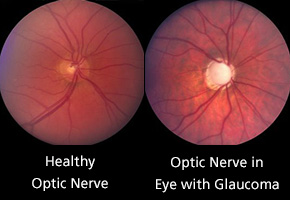

January is National Glaucoma Awareness Month. Glaucoma is a group of progressive eye diseases that damage the optic nerve, the bundle of nerve fibers that connects the retina, which converts light entering the eye into electrical signals, to the brain, which interprets those signals as visual images.
Continuing damage to the optic nerve from glaucoma can lead to vision loss. In fact, glaucoma is a leading cause of irreversible blindness in the US and the second leading cause of blindness in the world. It’s estimated that more than 3 million Americans are affected by glaucoma, and half of those people don’t know they have the disease. More than 120,000 people in the US are blind due to glaucoma.
The damage to the optic nerve from glaucoma is most often associated with an abnormally high pressure in the eyes called intraocular pressure (IOP). Many types of glaucoma have no warning signs. Symptoms typically don’t become noticeable until vision loss has begun. That’s why glaucoma is called a “silent thief of sight.”
High IOP is the result of an excess of fluid called aqueous humor, which bathes and nourishes the eye. Normally, the eye produces and drains this fluid at the same rate, maintaining a consistent pressure within the eye. An abnormality in the eye’s drainage system can cause the fluid to build up in the eye and increase IOP.
There are several types of glaucoma. The most common is open-angle glaucoma, which affects about 95 percent of people with the disease. With this type the drainage angle in the eye is open but the trabecular meshwork is blocked, which results in the build-up of fluid and an increase in eye pressure.
The drainage angle is located where the iris, the colored part of the eye, meets the cornea, the clear cap on the front of the eye. Fluid drains from the eye through this angle. The trabecular meshwork is a spongy material through which aqueous humor flows out of the eye.
Angle-closure glaucoma occurs when the iris bulges forward and physically blocks or narrows the drainage angle. Angle-closure glaucoma can be acute, where there’s a rapid and sudden increase in IOP, or chronic, which develops slowly over time.
Symptoms of acute angle-closure glaucoma come on quickly and may include eye pain, severe headache, blurry vision, rainbows or halos around lights, sudden vision loss, eye redness, and nausea or vomiting. Acute angle-closure glaucoma is a medical emergency. Seek help immediately if you notice any of these symptoms.
With normal-tension glaucoma, the optic nerve becomes damaged even though IOP is in the normal range. The reason this happens is unknown. The optic nerve may be highly sensitive or there may be less blood being supplied to the optic nerve due to another condition, such as atherosclerosis, the build-up of fatty deposits on the walls of the arteries that blocks blood flow.
Having high IOP is the main risk factor for glaucoma, but there are other factors that may increase your risk for developing the eye disease as well. Because glaucoma can destroy vision before any signs and symptoms are noticeable, it’s important to be aware of the risk factors. You may be at a greater risk for developing glaucoma if you:
- Are over age 60
- Are of African. Asian or Hispanic heritage
- Have a family history of glaucoma
- Have diabetes, heart disease, high blood pressure or sickle cell disease
- Have a thin cornea
- Are extremely nearsighted or farsighted
- Suffered an eye injury or underwent certain types of eye surgery
- Used corticosteroids, especially eyedrops, for an extended period of time
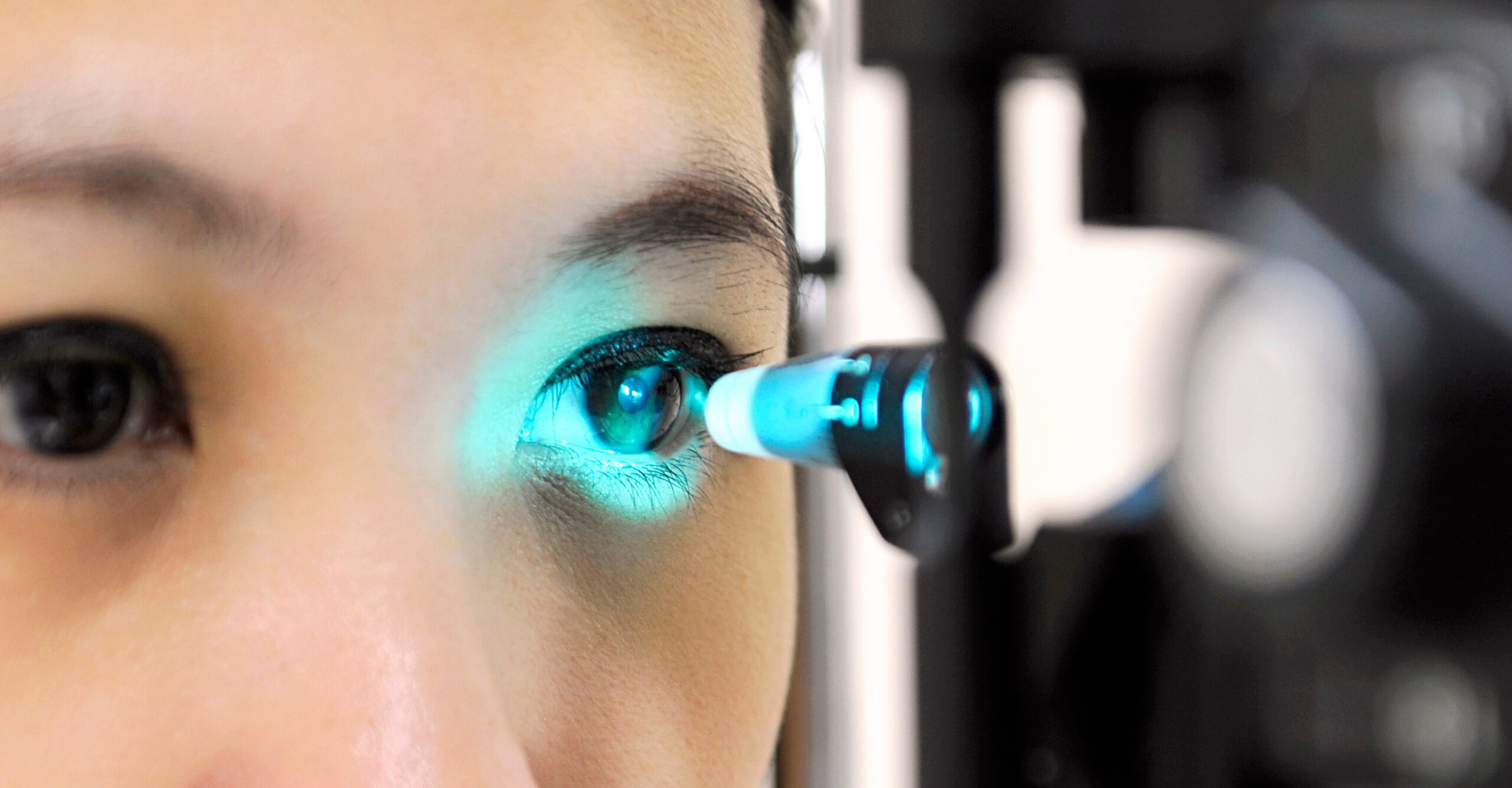
The best way to detect glaucoma in its early stages is through a comprehensive eye examination, including a dilated eye exam. Your doctor may also use certain tests, including the following:
- Tonometry measures eye pressure
- Optical coherence tomography (OCT) provides a clear image of the optic nerve to look for damage
- Visual acuity test checks for vision loss
- Visual field test looks for changes in your peripheral vision, which is often affected first
- Gonioscopy examines the drainage angle
- Pachymetry measures corneal thickness
Vision lost due to glaucoma is permanent. It cannot be restored. But treatment can slow down any additional vision loss. Glaucoma treatments include:
- Medication – Prescription eyedrops can help decrease eye pressure by improving how fluid drains from your eyes or by decreasing the amount of aqueous humor produced by your eyes. There are many types of eyedrops available that fulfill these functions.
- Laser treatment – This may be an option if you have open-angle glaucoma. The laser is used to open up blocked channels in the trabecular meshwork, allowing fluid to drain better.
- Surgery – Surgery is more invasive but can typically achieve better eye pressure control and do it faster than eyedrops or laser treatment. There are many types of surgery. Your eye doctor will select a surgery based on your specific type of glaucoma and its severity.
Don’t let glaucoma steal your sight. Routine eye exams can catch this disease in its early stages, and prompt treatment can help slow the progression of glaucoma and save your vision. Ask your eye specialist how often you should have your eyes examined.
iFoundMyDoctor.com article by Patti DiPanfilo


Leave a Reply